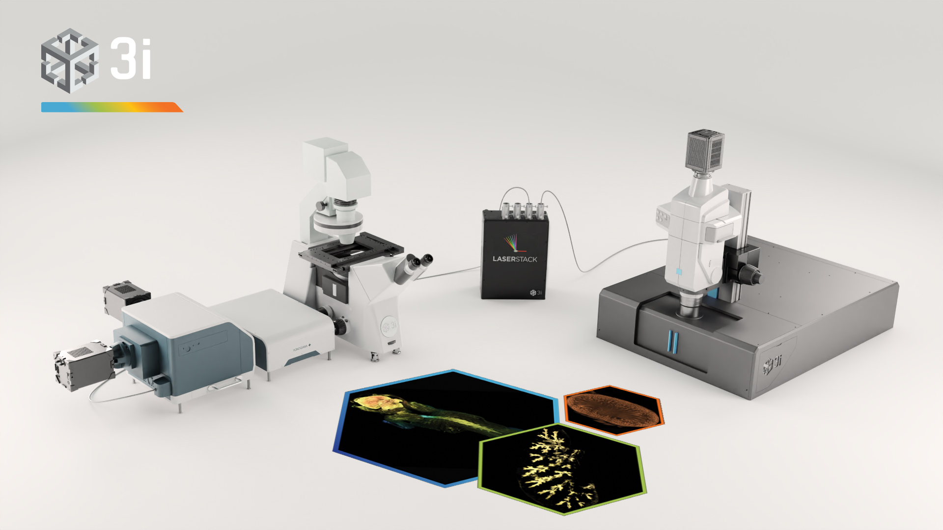
Clearing tissue is now a powerful and accessible technique for studying thicker samples in greater detail. The recent proliferation in tissue-clearing reagents and methods has made the technique more effective and more reliable over a greater range of samples from tissues to organs to whole animals. Once a suitable tissue-clearing regime has been established for a sample the following consideration is what image acquisition modality to use? Two potential options are a spinning disk confocal and a lightsheet microscope, both of which we will present in this workshop. Some of the parameters of a sample or the feature of interest will clearly direct this decision; for example, imaging a whole cleared mouse would typically preclude a spinning disk confocal and imaging a cleared tissue sample at super-resolution would typically preclude lightsheet microscopes. When imaging cleared specimens multiple factors need to be taken into consideration, foremost is a careful evaluation of the volumetric data such that it meets resolution requirements and is an accurate 3D representation of the sample. Other considerations are practical including: safety and ease of sample handling, ease of capture and duration of data acquisition. In this workshop we present our Marianas SDC inverted microscope system featuring a CSU-W SoRa super-resolution spinning disk confocal and our Cleared Tissue LightSheet (CTLS) microscope system and explore cleared tissue samples that could be imaged by each modality and compare the benefits and limitations of each technique.
Workshop 1
WED 8 JUNE 2022, time: 13:30 - 14:30
Click on the images to open the full image view!
3i - Intelligent Imaging Innovations
Room: Logi 2
A comparison of spinning disk confocal and lightsheet when imaging at different scales
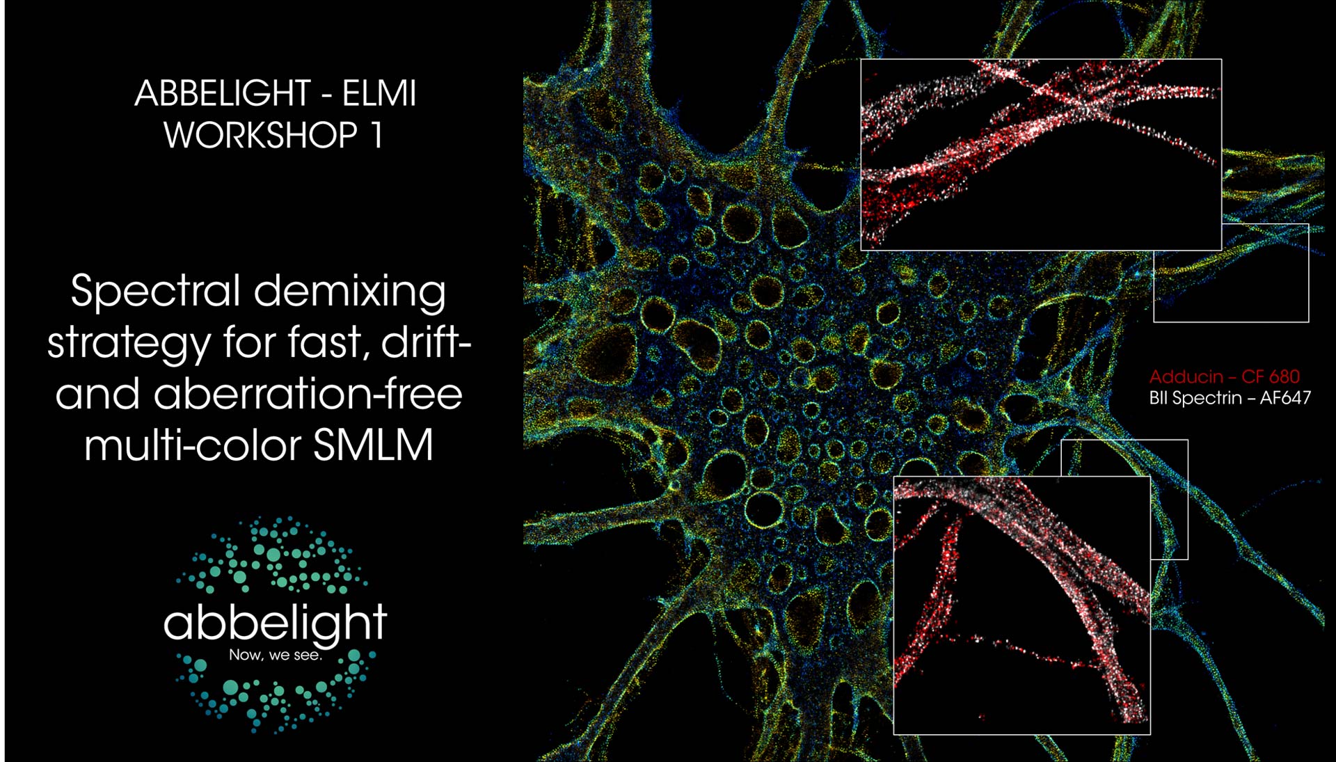
Abbelight
Stand 36b
Spectral demixing strategy for fast, drift- and aberration-free multi-color SMLM
Different spectral demixing methods have been proposed by the SMLM research community: diffractive-based approaches offering a spectrum analysis of each single molecule in addition to its spatial location (Zhang, Z. et al. 2015) and ratiometric approaches (Bossi et al. 2008; Testa I. et al 2010; Baddeley et al. 2011; Lampe et al. 2012; Winterflood et al. 2015, Diekman et al. 2020) which are simpler and more robust. As simplicity and data robustness are two major focus of abbelight, the spectral demixing strategy adopted relies on the ratiometric method.
During this workshop, we will present the imaging by sprectral demixing with the SAFe 360 which the principle is to split the emission fluorescence into two distinct detection channels thanks to a long pass dichroic beamsplitter. By using fluorophores emitting within the same wavelength range, their partially overlapping emission is then spectrally separated by the dichroic and imaged onto two detection scientific-grade cameras (the choice of the dichroic is linked to the fluorophores and the SMLM strategy employed). The Point Spread Function (PSF) of each fluorophore can be retrieved on both images provided by the two cameras and the measured photon numbers on each camera will be related to the spectral separation of the fluorophore.
Schedule:
10 min – Introduction to Spectral-demixing
15 min – Acquisition of single-color image for control
5 min – Short break to change sample
20min – Simultaneous Multi-color Acquisition using Spectral-demixing
15 min – Summary of the workshop & Questions
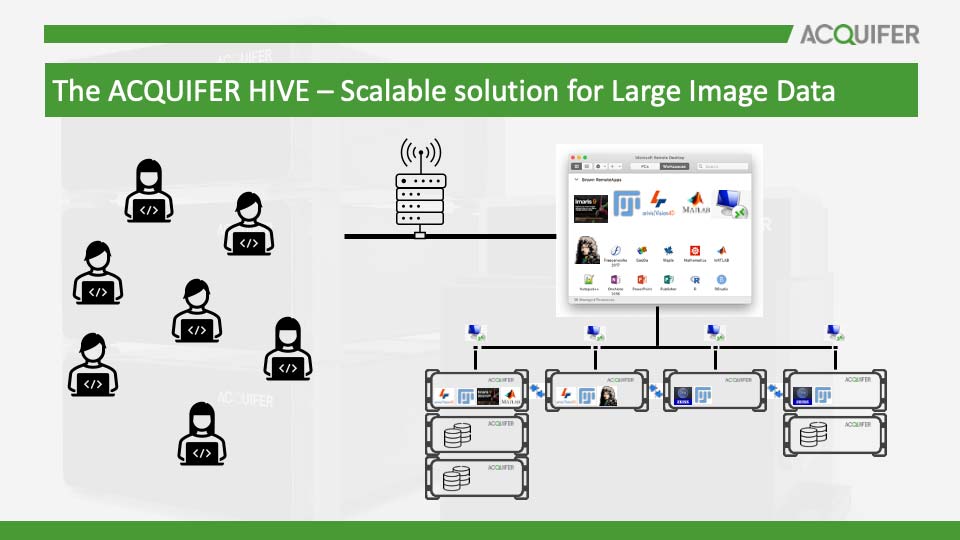
ACQUIFER Imaging GmbH
Room: Gallery 1
The ACQUIFER HIVE - Solution for Large Image Data
Modern microscopy techniques acquire an ever-increasing amount of multidimensional image data. 3D microscopy techniques, such as selective plane illumination microscopy (SPIM), easily generate multiple terabytes of raw image data in a single experiment that require tailored solutions for interactive or automated analysis.
The ACQUIFER HIVE is the established solution for efficient and safe management and storage of large image data without USB drives or network copy. The HIVE allows fast access to acquired data for swift processing and flexible analysis workflows. It is designed as a modular platform that is configured for your specific applications and will grow with your needs.
In this workshop, we will introduce the concepts of HIVE workflows, and we will present new solutions to expand existing HIVE installations to serve a higher user base and larger projects. Multi-GPU and Multi-HIVE solutions are outlined. Besides scaling in processing, concepts about data safety with large storage pools are discussed.
As part of the workshop, we offer RDP access on the centralized multi user system HIVE for testing. We encourage participants to bring their own laptops.
Please refer to our website at www.acquifer.de or call us at +49 (6221) 435 2000 for more detailed information on ACQUIFER products.
ARGOLIGHT
Stand 9
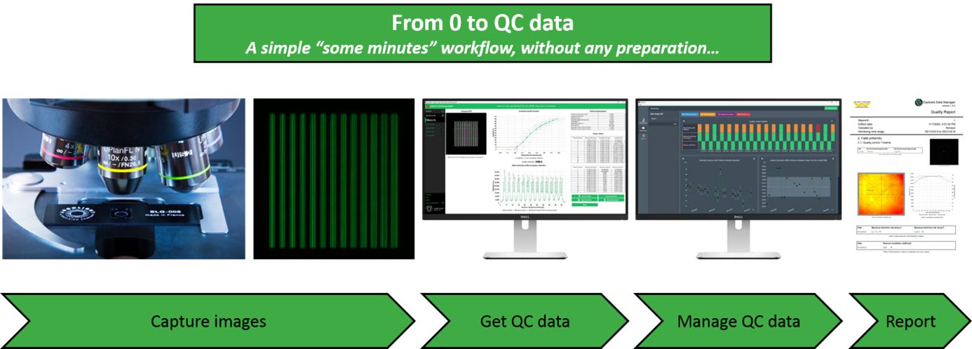
Obtain and manage quality control data on your microscopes
One of the core facilities’ duties is to provide end-users, usually researchers in life sciences, a fleet of microscopes at a level of performance compatible with their experiments. This is not an easy task because the performance of such devices tends to fluctuate or deteriorate over time for many reasons: misusing, aging, environment fluctuations, etc. This is especially true for high end imaging systems such as confocal or super resolution microscopes. Having access to a unique, reliable, and easy-to-use device to ensure microscopes’ performance would certainly make easier this tedious task.
To get quantitative and reproducible data, assessing the performances of fluorescence microscopes is a prerequisite before any imaging campaign. For example, system co-registration accuracy should be evaluated before any co-localization study; System field uniformity and intensity response before any study where intensity in the image matters; Spatial resolution before any study aiming at counting objects close to each other, etc.
Three years after our last presence at ELMI, we are happy to come back and present during this workshop the novelties we have developed in the meantime: improved hardware products, new analyses from 3D patterns, new analyses related to non-Argolight products, improvements in user interface and user experience, etc. Above all, the presentation aims to show how the quality control of fluorescence microscopes can be standardized with Argolight software solutions, and how the generated quality control data can be managed and centralized for later reporting.
Bruker
Room: Goto 32
Imaging across scales – from single particle to whole living and cleared organisms
In this workshop we will demonstrate Bruker super-resolution and light-sheet microscopy solutions (Luxendo SPIM family).
The SPIM demonstration will cover different sample types and potential applications ranging from life imaging of delicate live specimen such as organoids and embryos (mouse, zebrafish etc. ) to whole in-toto imaging of large cleared samples such es entire adult mice. We will be showing our 3D Super-resolution system, the VXL with patented 3D technology suitable for STORM and PALM, along with an integrated
microfluidics system for highly multiplexed, DNA-PAINT research.
Workshop 1/4 : TruLive3D SPIM – light-sheet microscopy for live samples
Workshop 2/5 : VXL – 3D super-resolution microscopy
Workshop 3/6 : LCS SPIM – light-sheet microscopy for cleared samples
Image:
Top, left image: live zebrafish embryo H2A::GFP recorded on Luxendo MuVi SPIM LS. Top, right image: Mitochondria stained for TOM20 with Alexa 647 and imaged with STORM. Bottom, left image: cleared adult mouse with GFP labeled nerves and recorded on Luxendo LCS SPIM. Bottom, right: Cleared YFP expressing transgenic mouse brain recorded on Luxendo MuVi SPIM CS

Carl Zeiss Microscopy
Room: Goto 33
Discover the evolving technology of Lattice Lightsheet Microscopy
The launch of the ZEISS Lattice Lightsheet 7 inverted platform in 2020 was an important milestone for lattice lightsheet microscopy. This platform was designed for use by the non-expert so no alignment or technical know-how is needed to capture data. This has led to wide adoption of the Lattice Lightsheet 7 and it has now emerged as one of the key solutions for long term live imaging in the core facility. Further expansion of the Lattice Lightsheet 7 platform to more capability both in acquisition and analysis to assist in scaling experiments to higher throughput now further extend the imaging possibilities.
This workshop will focus on lattice lightsheet technology and will introduce the new capabilities that the ZEISS Lattice Lightsheet 7 offers. You will see the full workflow in action from capturing the data through to segmentation and quantitative analysis of the resulting datasets using arivis Vision 4D.
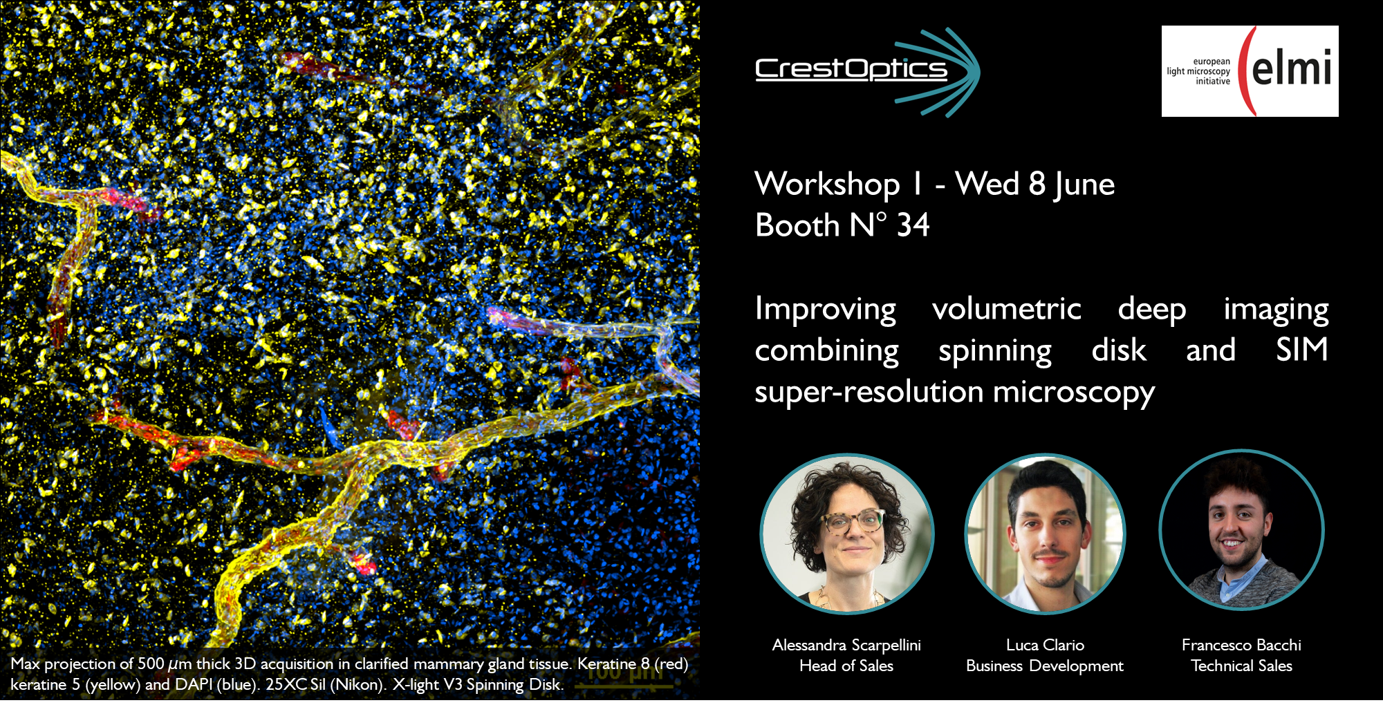
CrestOptics SpA
Stand 34
Improving volumetric deep imaging combining spinning disk and SIM super-resolution microscopy
Fast and highly resolved volumetric microscopy is essential to examine various biological aspects in their complexity, ranging from whole animal imaging, to live phenomena, to thick samples to image at the different depth. During this workshop we demonstrate how X-light V3 spinning disk and DeepSIM add-on can be combined to work with deep samples of thickness comparable to those normally used in CF microscopy. This means that more meaningful data can be obtained from native heterogeneous complex (over 100µm thick) samples using routine preparation protocols.
#deepimaging #clearedtissues #spinningdisk #SIM
Cytena
Stand 8
From 96-wells to single cells: Introduction to high content live cell assays on Livecyte
Characterising, comparing, and ultimately understanding, differences in dynamic live cell behaviour is critical in many fields, from cancer research to regenerative medicine.
In this workshop, we explore new ways to characterise cell behaviour, enabled by the Livecyte Kinetic Cytometer system. We look at how simple label-free assays, in 96-well plates, can be used to independently measure cell growth and cell proliferation, automatically characterise both random and sheet cell migration and identify other phenotypic changes. We also show Livecyte’s correlative fluorescence capabilities, and how these measurements unlock additional powerful new insights.

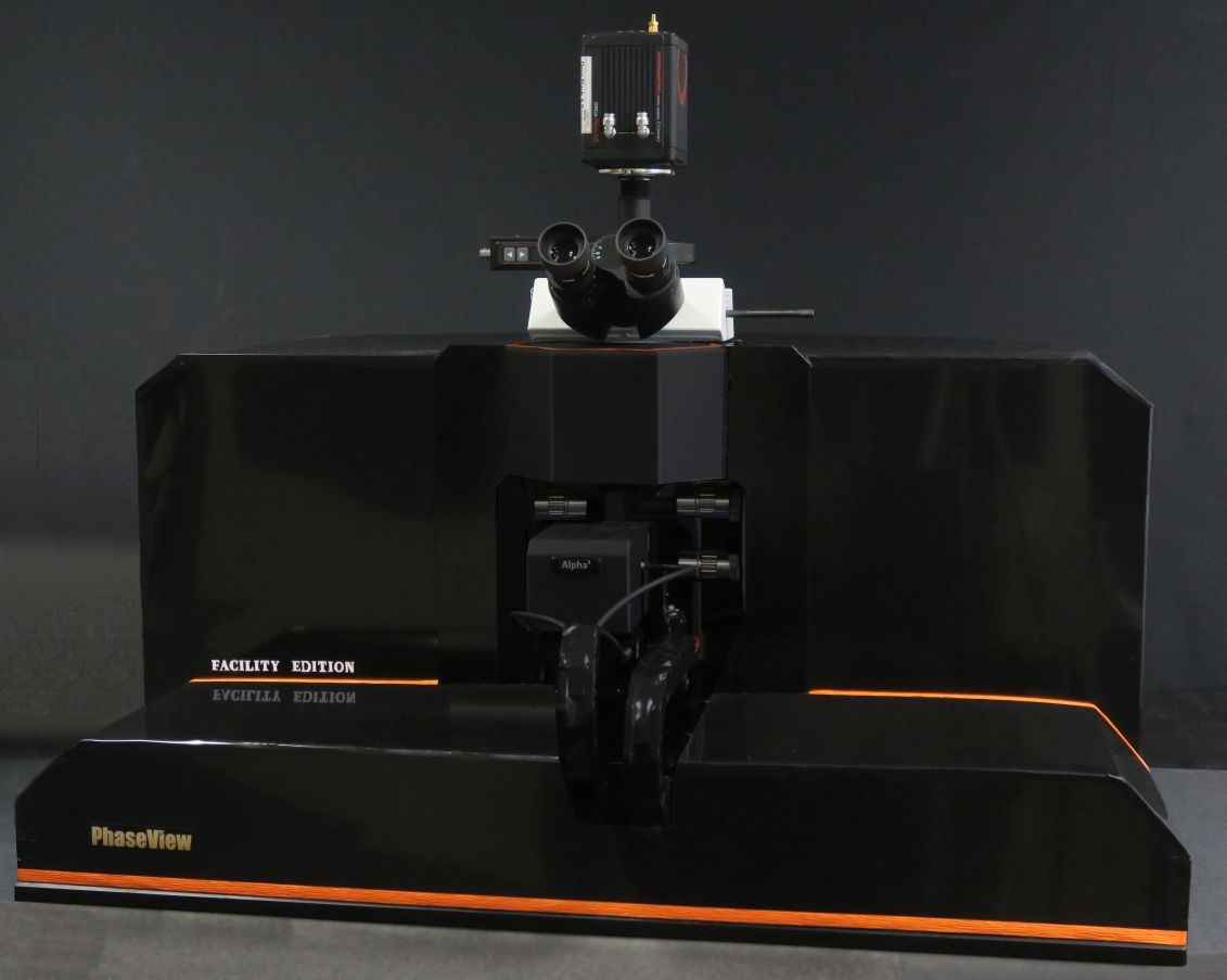
EVIDENT/OLYMPUS
Room: Bäkkäri
Ultra-thin light sheet microscope with automated multi-scale imaging capabilities
| Presenter: Irina Rakotoson / PhaseViewAbstract: The new fully automated Facility Edition light sheet microscope combines key technologies to deliver optimal image quality with an ultra‐thin light sheet thickness while offering an unrivaled user experience from fast sample screening to high-res image acquisition of whole cleared specimens. |
ibidi GmbH
Stand 29
ibidi Imaging Chambers and Surfaces
High-resolution microscopy of cells requires imaging chambers that are specifically tailored to your application.
Are you interested in learning about…
• … easy to handle all-in-one solutions for live cell imaging?
• … the advantages of using channel slides for immunofluorescence stainings?
• … solutions for 3D cell culture and spheroid imaging?
Join our workshop and find the answers to these questions!
First, we will introduce a variety of ibidi chambers with different geometries and surfaces and help you to choose the products that are best suited for your assay. This will be followed by a hands-on-training where you get the chance to test a selection of our labware for 2D and 3D cell culture and imaging.

Leica Microsystems CMS GmbH
Room: Teatro
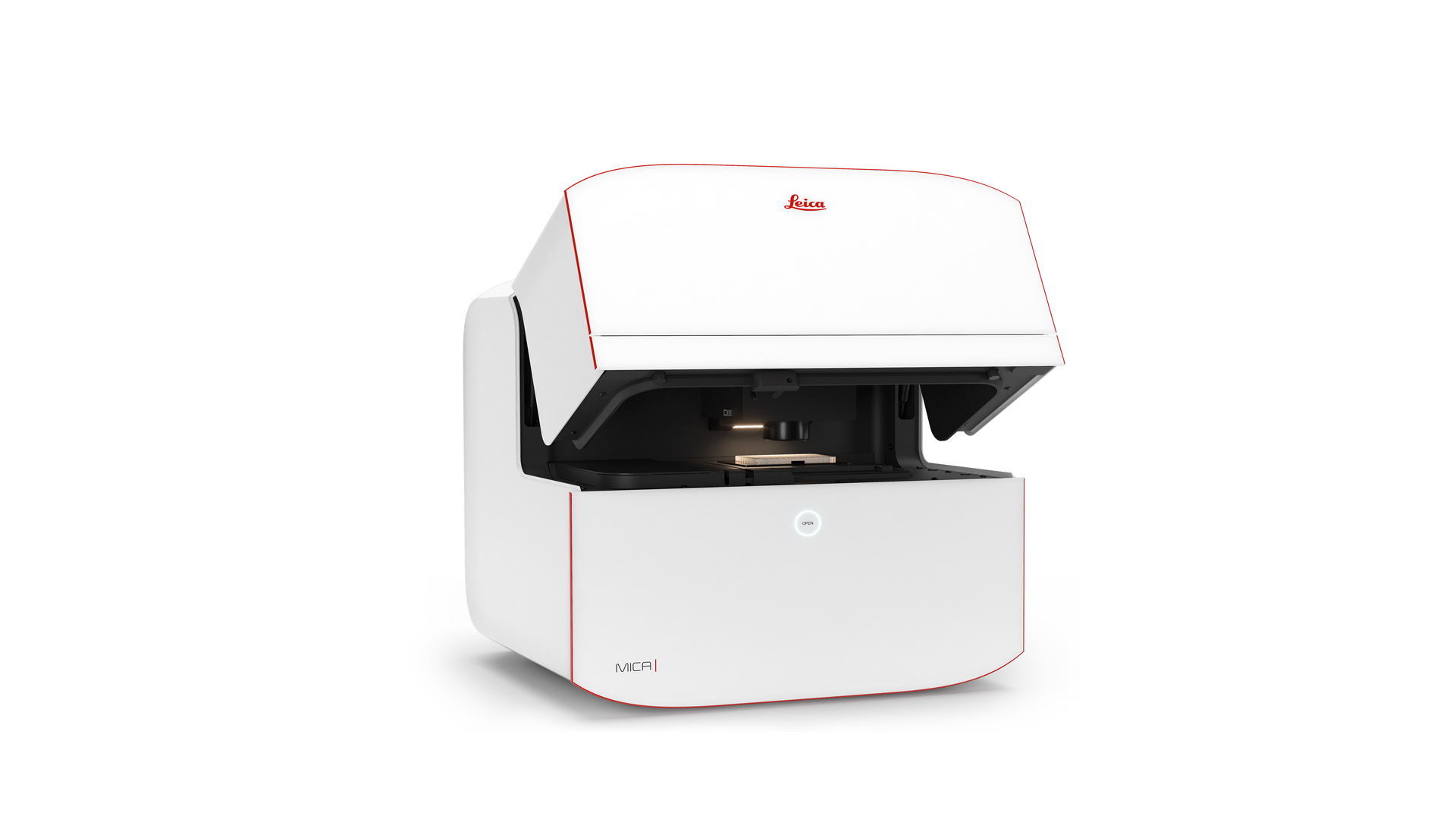
Enter the Microhub Era - Meet Mica!
Experience Mica in action and learn how its sample-centric operation allows straightforward access to imaging for all users .
See how MICA dramatically reduces the time-to-image and training time for non-imaging specialists.
Learn how our new FluoSync technology detects four fluorophores simultaneously, in both widefield and confocal!
Discover how the whole workflow from setting up an imaging experiment to the AI powered extraction of data has been radically simplified.
See all this working directly on samples and specimens you see every day – from multi-well plate assays to large tissue sections to model organisms – all on Mica!
Cutrale, F., Trivedi, V., Trinh, L. et al. Hyperspectral phasor analysis enables multiplexed 5D in vivo imaging. Nat Methods 14, 149–152 (2017). https://doi.org/10.1038/nmeth.4134
·Hsiao Chiang, Daniel Koo, Masahiro Kitano et al. HyU: Hybrid Unmixing for longitudinal in vivo imaging of low signal to noise fluorescence, 12 January 2022, PREPRINT (Version 1) available at Research Square [https://doi.org/10.21203/rs.3.rs-1073331/v1]
White paper: https://webcdn.leica-microsystems.com/fileadmin/global/products/Supercharge-the-way-you-work/White_paper_FluoSync_MC-0003306.pdf
Miltenyi Biotec B.V. & Co. KG
Room: Gallery 2
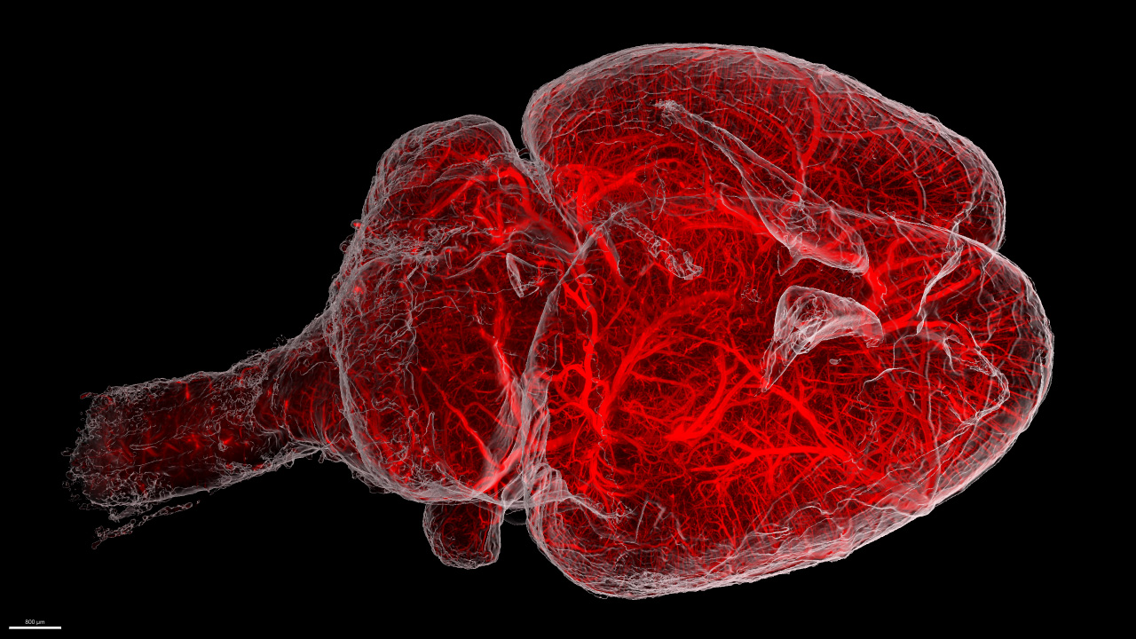
Dive deep into your sample – the Miltenyi 3D imaging workflow
Visualizing the three-dimensional architecture of complex organisms while analyzing biological processes in a whole-organ fashion are becoming the new standard in research. To provide a complete, smooth, and hassle-free 3D imaging workflow, Miltenyi Biotec covers this entire process offering solutions for sample staining, clearing and imaging. In this workshop, we will guide you through the most important steps of sample preparation, including a live demonstration, and will demonstrate how easily high quality 3D images of large samples can be acquired with our cutting-edge light sheet system, the UltraMicroscope BlazeTM.
Nikon Europe BV
Room: Move 1
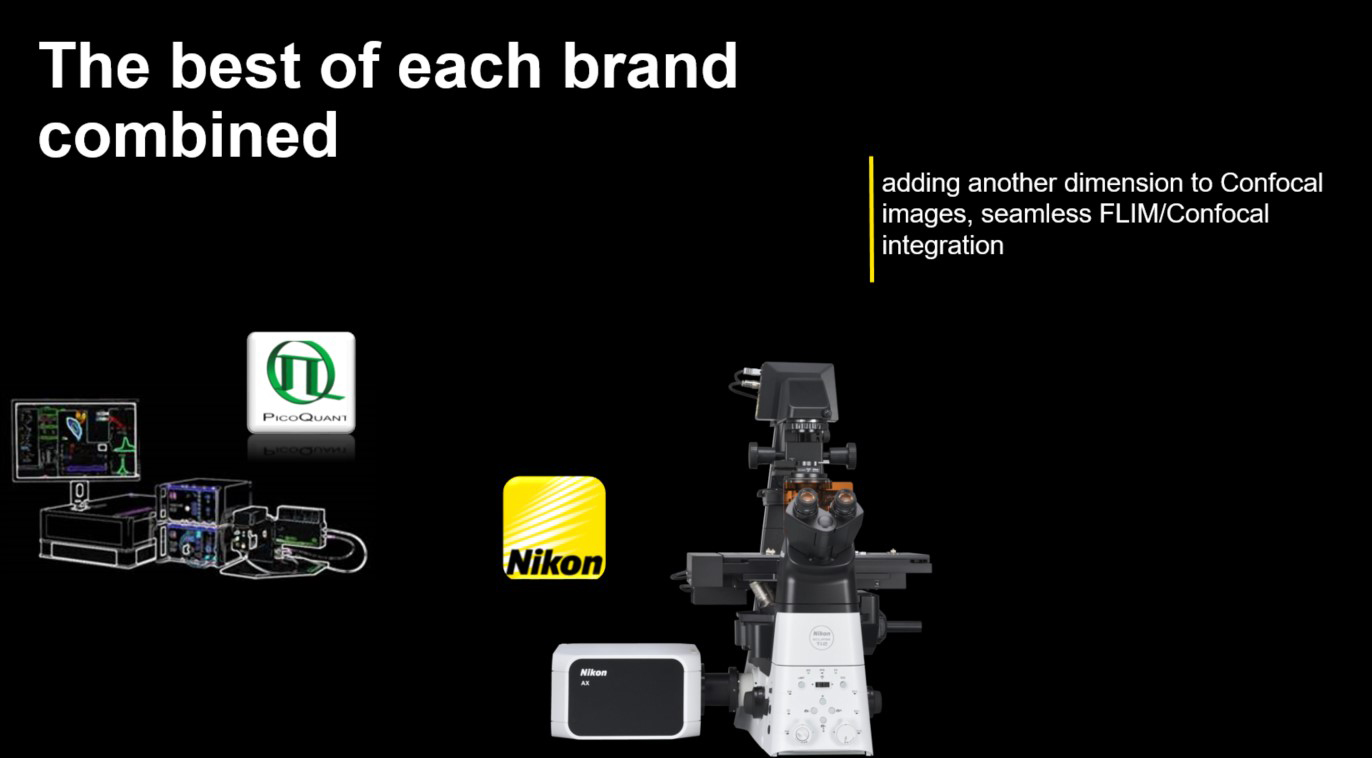
Expand to lifetime imaging: A seamless integration of AX point scanner confocal and FLIM
With AX’s new capabilities and easily managed lifetime experiment combination, we add an extra dimension to confocal microscopy.
In this workshop, you will find out how one can acquire and visualize FLIM data with the help of NIS-Elements. Discover a quick and intuitive basic analysis package directly included in NIS-Elements: Large image, Z stack, automated complex acquisitions, and basic analysis. Easy export of FLIM image to Symphotime within one click for more in-depth is at reach.
There is much more to discover, so join us and learn how AX/FLIM provides seamlessly integrated lifetime acquisition with unprecedented resolution.
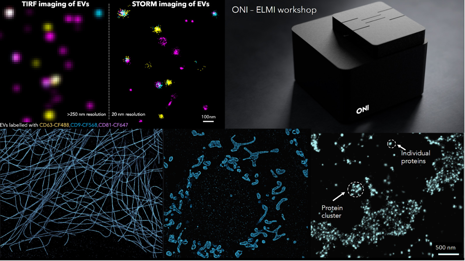
ONI
Room: Move 2
Every Molecule Counts - the ONI super-resolution platform just got even better
Studying spatial protein organization or protein copy number, clustering and mobility at the molecular level is extremely important from both a diagnostic and treatment perspective. Looking at cellular structures beyond the diffraction limit with a resolution of 15-20 nm with robust quantification has been enabled with Single-molecule localization microscopy (SMLM) like STORM or PALM. ONI has created the world’s first desktop super-resolution microscope, the Nanoimager, which is designed to operate on a standard lab bench and has a footprint smaller than a piece of A4 paper, with the most intuitive and simple software interface, making it also accessible to researchers not specialized in high end microscopy techniques with one major goal: make it accessible and simple to use.
It is really important to enable users to extract quantitative information from their localization-based images. We have done this by designing a cloud-based analysis software called CODI (COllaborative DIscovery). CODI helps users analyze their single-molecule data by extracting quantitative information on protein clustering, cluster size and area, cluster density, and so much more. It enables all researchers to upload their data, share or collaborate with other scientists, whilst gaining meaningful insights about their data.
During this workshop we will perform a two-color STORM on mitochondrial samples (TOM20 and HSP60) and reveal the sub-mitochondrial compartmental organization with 20 nm resolution. The aim is to introduce SMLM imaging as a powerful tool for rapid and multiplexed characterization of cellular structures along with a visual and quantification analysis platform CODI developed by us. We will follow up the workshop with an open discussion on the wide-ranging applicability of the Nanoimager in different biological applications encompassing the field of immunology, neurobiology, cell biology and cancer.
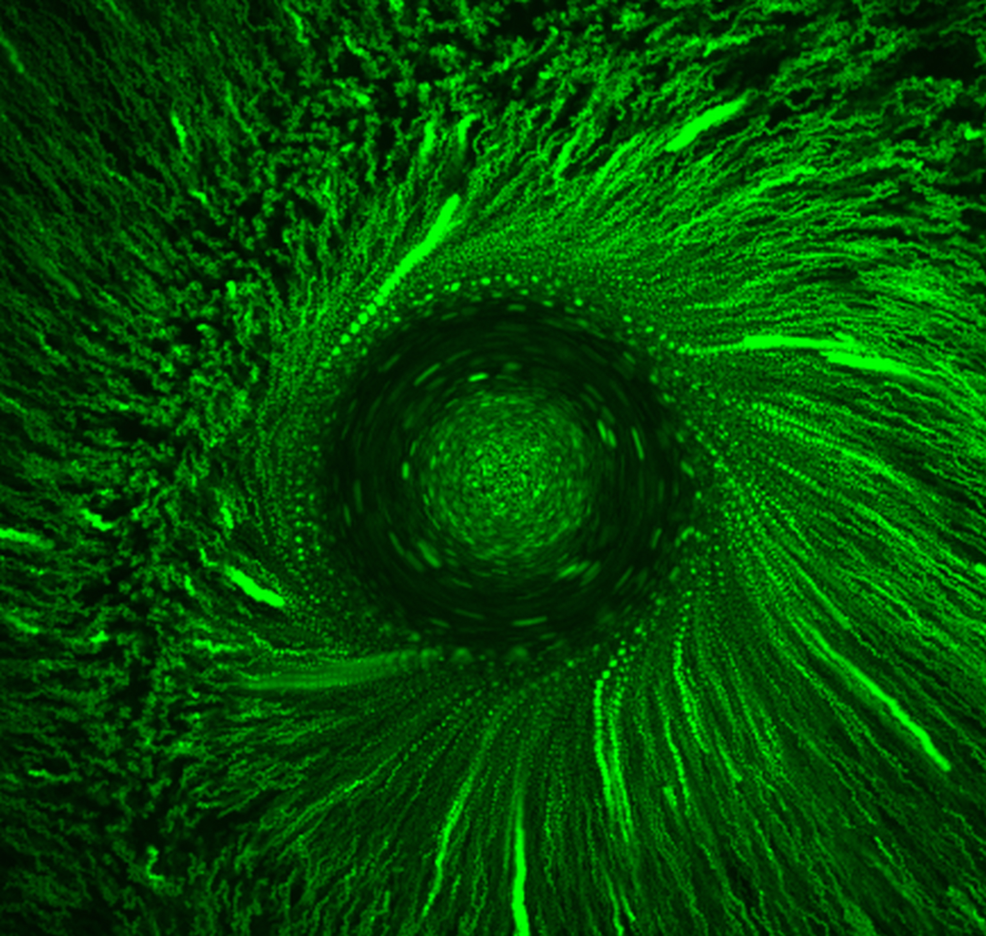
Prospective Instruments
Room: Goto 31
First turnkey Multiphoton Microscope!
Bring your own sample to the conference to image it with Prospective Instruments MPX series Two-Photon Microscope. Contact us if you would like to take this opportunity.
Of course, we will also bring a selection of samples to image.
Rapp OptoElectronic GmbH
Stand 35
FLUCS: Micro flow photomanipulation
FLUCS photomanipulation is a non-invasive, all-optical method based on thermoviscous flow that allows the microscopist to induce precise microscopic flow fields inside living cells or microfluidic chambers.
During the workshop, we present RAPP’s FLUCS add-on optical module that is compatible with all standard inverted microscopes, we explain the working principle of FLUCS technology, we discuss a variety of recent applications of this novel technology in the life sciences, and we provide guidance, to optimally exploit the technology for your experiment.
www.rapp-opto.com/products/photomanipulation-systems/flucs-micro-flow-photomanipulation/
Thermo Fisher Scientific
Room: Goto 30
Explore large multi-channel and time series data with Amira Software and the new Xplore5D extension.
During these workshops you will discover, through a step-by-step demo, the recently added functions of the Thermo Scientific™ Amira™ Software’s new Xplore5D extension, which offers data conversion and compression with immediate visual feedback, allowing you to quickly interact with and communicate on large 3–5D data.

TissueGnostics GmbH
Room: Logi 1
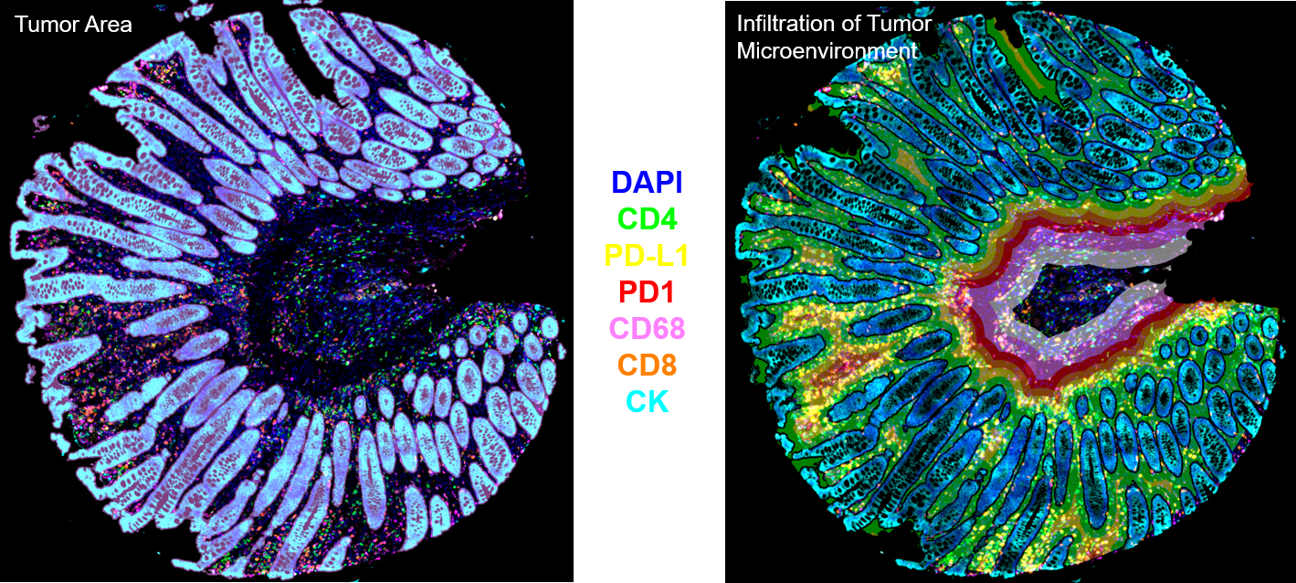
Spatial Tissue Cytometry - Using Precision Microscopy with Artificial Intelligence
Tissue Cytometry permits to determine the in-situ phenotype of cells as well as histological entities, like glands, vessels or tumor foci. Applications include but are not limited to the exploration of the cellular/tumor microenvironment and/or the spatial organization of cellular subpopulations, assessment of different bone structures, quantification of blood vessels and neovascularization as well as analysis of samples in multiplexing or multispectral mode.
Earlier attempts to analyse single cells in tissue have mostly been subject to visual estimation, or – at best – to manual counting for decades. Hence, experts usually had the choice of the “least of evils” between guessing and endless (manual) counting. In (tumor) immunology, infiltrating inflammatory cells need to be phenotypically characterized on a quantitative basis. To better understand the function of inflammatory cells in tumor development, type and number of inflammatory cells and their proximity to glandular/tumor structures have to be analyzed in-situ and correlated with disease state. Using TissueFAXS™ Cytometry the time-consuming and error-prone human evaluation of stained histological sections can be approached with an observer-independent and reproducible technology platform, offering a high degree of automation, paired with user interaction at relevant points of the analytical workflow. TissueFAXS Cytometry is the “one-stop shop” for all kinds of microscopic analysis of tissue sections, organoids and adherent cell cultures.
Interherence
Room: Main Auditorium
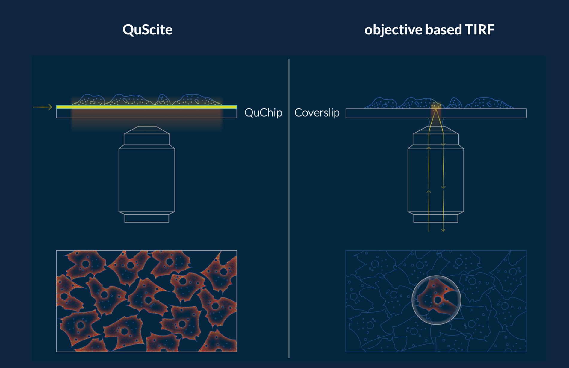
Reproducibility through precision: Chip-based technologies for precise control of sample temperature and illumination in high- and super-resolution microscopy.
Biological systems are complex and many environmental parameters affect their behavior. The chemical conditions of the sample volume, including pH and salt concentration, are often well considered when designing experiments in high and super-resolution microscopy. However, control over the physical conditions such as temperature or the amount of light deposited in the sample volume are often not well characterized and might differ from setup to setup. The imprecisions mainly arise due to the lack of appropriate sensor that can be placed close to or directly in the sample volume. Integrated systems, which are in direct contact with the sample, can help to improve reproducibility in biophysical studies of living cells, proteins or DNA and make interpretation of the acquired data easier.
Measuring and controlling the temperature of small sample volumes, as is often the case in high and super resolution microscopy, is technological challenging. In the first part of this talk, we will review state of the art approaches for temperature management in light microscopy, analyze their strengths and weaknesses and talk about our solution, VAHEAT. We will highlight the importance of controlling the temperature in biophysical studies based on recent research covering the fields of single molecules studies, live cell imaging and colloid chemistry.
In the second part of the talk, we will focus on illumination conditions specifically in total internal reflection (TIR) microscopy. After analyzing the limits of current technologies, we take the opportunity to pre-launch QuScite – our device for TIRF microscopy on a chip. This waveguide TIR system relies on photonic integrated circuitry (PIC) and allows to separate the excitation from detection path, which opens up completely new pathways for studying single molecules and processes in cell membranes. QuScite’s unprecedented field of view in the range of square millimeters combined with its ease of use even in complex workflows should allow everyone, on any microscope to perform high sensitivity TIRF measurements under reproducible conditions.
Note: This presentation will be interactive. You will have the opportunity to see and test demo systems on site.
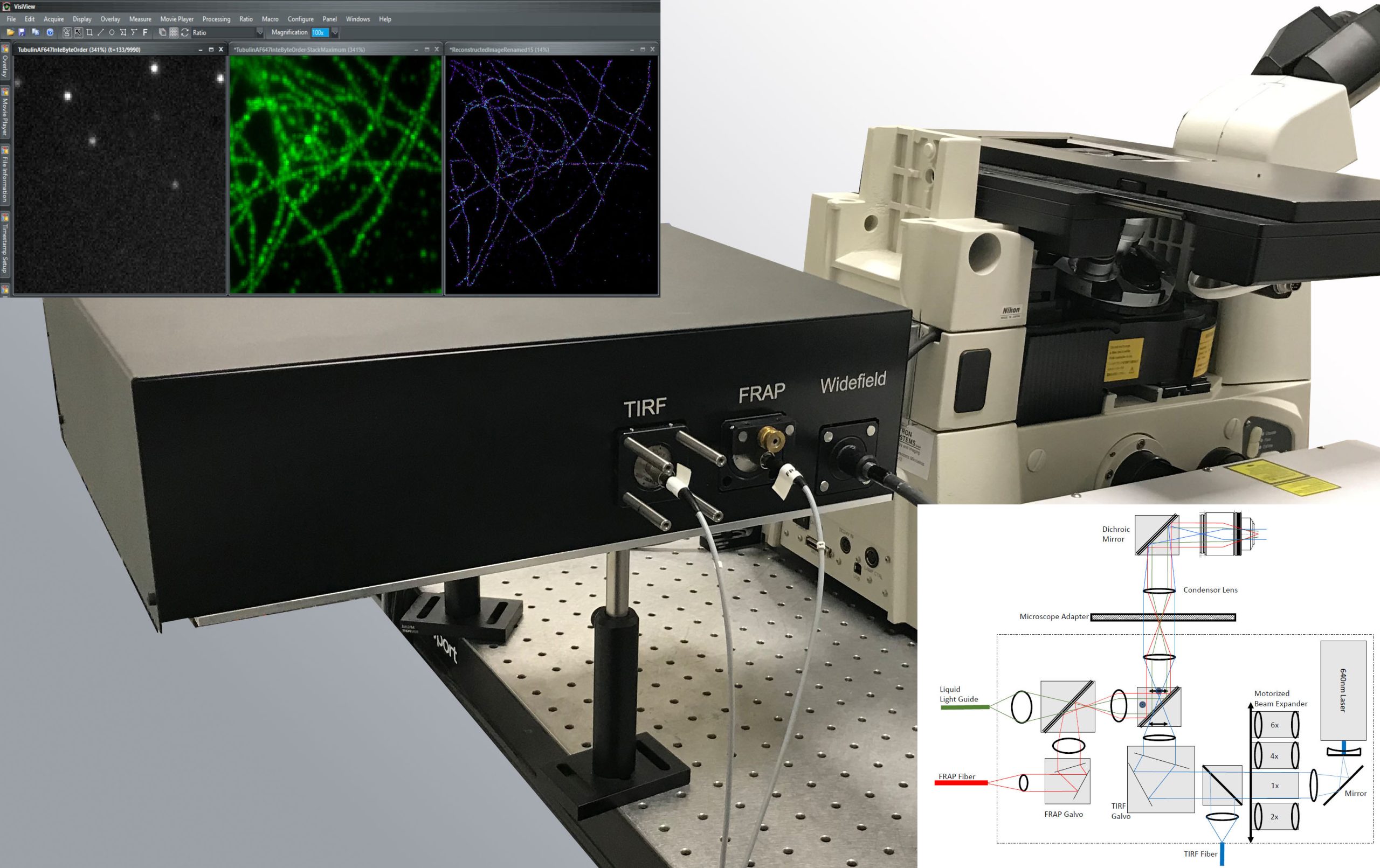
Visitron Systems GmbH
Stand 27
VisiTIRF Orbital RingTIRF Technology for STORM or VisiScope Spinning Disk Microscopy
This workshop will give the listener an overview of new developments in RingTIRF technology, new features in VisiView Imaging Software and highly accurate experiment control by ViRTEx.
2. ORBITAL Ring-TIRF Technology
The VisiTIRF-ORBITAL is a compact and powerful high speed 2D galvo driven spinning Ring-TIRF laser illumination system. It offers a large and evenly illuminated field of view to enable applications such as single molecule tracking or SMLM – Single Molecule Localisation Microscopy for superresolution imaging. Full 360 degree positioning by free circular diameter or elliptical trajectory at the back focal plane of the high aperture TIRF objective offers illumination with minimal fringes or shading gradients